ISO9001 Certified Professional Manufacturer & Supplier of Optics
+86-0431-87911611 admin@ytoptics.com
Contact us
-
 Email: admin@ytoptics.com
-
 Tel:86-0431-87911611
-
 Add: 2# Automotive Innovation
Jilin Province, China
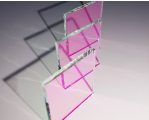
Fundus Camera Filter
Fundus angiography is the process of injecting fluorescent contrast agent into the patient through the vein, and at the same time using a special filter combination of fundus camera for fundus photography, dynamic observation of the whole process of contrast agent circulation in the fundus of the retina and choroid, which can reflect the damaged state of the retinal barrier in the eyes of the living person, and dynamically capture the physiological and pathological conditions of the retinal blood vessels to the capillaries. With the help of fundus angiography results, clinicians can diagnose and deal with common fundus diseases such as diabetic retinopathy, age-related macular degeneration, hypertensive retinopathy, venous obstruction, and glaucoma optic disc, retinal nerve fibre layer analysis, etc., as well as observe changes in the lesions and evaluate the treatment effects. With fundus fluorescence angiography as a special examination method, the diagnosis of patients' fundus disease is changed from subjective observation to an objective scientific identification, that is to say, from the static observation under the funduscope to a dynamic examination, which can discover the lesions that cannot be detected by the common fundus examination. Another important role of fundus fluorescence angiography is to be used for pre- and post-treatment examination of fundus diseases and to determine the efficacy of treatment. For example, retinal vein occlusion and diabetic retinopathy should be treated with fluorescence angiography before laser photocoagulation treatment to understand the lesion condition and formulate a precise treatment plan.
Share this:
PREV : Principle of the Chromatic Prism NEXT : Difference between concave and convex lenses
TALK TO US 86-0431-87911611
Call us now!
86-0431-87911611Call us now!

ONLINE CHAT
 2433808388